

FEATURE
FEATURE1 High-resolution images obtained with the FE-SEM.
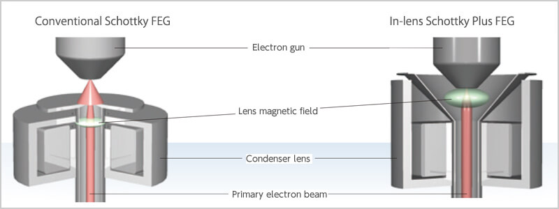
In-lens Schottky Plus Field Emission electron Gun (FEG)
The In-lens Schottky Plus FEG has realized improved brightness as a result of integration of the electron gun and low-aberration condenser lens. With this FEG, generated electrons can be efficiently focused, enabling probe currents on the order of a few pA to several tens of nA even at low accelerating voltages. High-resolution observation is easy: there is no need to switch the objective aperture for tasks from fast elemental mapping to EBSD to SXES analyses.
Hybrid Lens (HL)
The JSM-F100 comes with JEOL's electrostatic/electromagnetic field superposed objective lens, Hybrid Lens (HL). This powerful lens enables observation and analysis of any specimen, including magnetic and insulating materials at high spatial resolution.
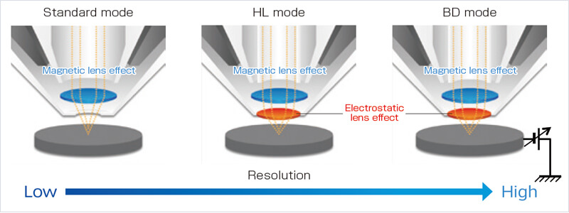
- ※BD(Beam Deceleration):
- Applying a bias voltage of up to −2 kV to the specimen stage enables deceleration of the incident electron beam just before the specimen. This function improves the spatial resolution and signal-to-noise (S/N) ratio at low accelerating voltage.
The JSM-F100 FE-SEM permits observation of nanostructures. Selecting observation conditions and detectors suitable for your applications enables you to acquire characteristic SEM images from a variety of specimens.
-
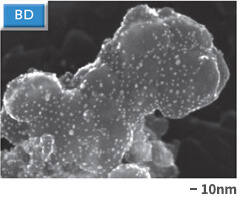
Specimen: Pt nanoparticles on carbon,
Accelerating voltage: 20 kV, WD: 2 mm,
Observation mode: BD, Detector: UED -
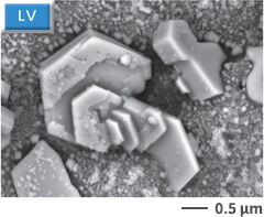
Specimen: Electric lamp lead,
Accelerating voltage: 10 kV, WD: 6 mm,
Observation mode: LV, Detector: LVBED -
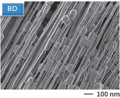
Specimen: Seal tape,
Accelerating voltage: 0.5 kV, WD: 2 mm,
Observation mode: BD, Detector: UED -
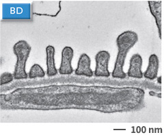
Specimen: Ultra-thin section of mouse glomerulus,
Accelerating voltage: 5 kV, WD: 4 mm,
Observation mode: BD, Detector: RBED (contrast reversal)
- ※ LV (Low Vacuum function) *Option:
- The Low Vacuum function allows simple observation and analysis with no conductive coating for insulating materials.
FEATURE2 Highly Innovative Operability
With optical imaging, SEM imaging and EDS analysis integrated,
measurement throughput will be dramatically improved.

FEATURE3
SMILE VIEW™ Lab
Automatic linkage of optical and SEM images and EDS results
The JSM-F100 offers connectivity with SMILE VIEW™ Lab, the new data management system used for JEOL's analytical instruments. It is useful for data management, analysis and report generation.

- ※ Aperture angle Control Lens (ACL):
- The aperture angle control lens (ACL), located above the objective lens, automatically optimizes the aperture angle of the objective lens over the whole current range (1 pA to 300 nA). Even when the probe current is increased, the ACL suppresses the spread of the incident electrons to continuously maintain the smallest possible probe size. The ACL enables smooth operation at any level of probe current, from high-resolution observation to analysis requiring a high probe current.
NEW FUNCTION
NEW FUNCTION 1
In-built AI for SEM
LIVE-AI Filter (Live Image Visual Enhancer – AI: LIVE-AI)※ Option
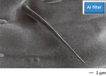
JEOL has incorporated the LIVE-AI (Artificial Intelligence) filter for higher quality of live images. Unlike image integration processing, the LIVE-AI filter can display a seamless moving live image with no residual image. This unique feature is very effective in searching for the observation area and for focus and astigmatism correction.
Comparison of normal mode and LIVE-AI filter
-
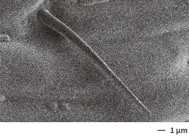
Normal mode
-
With LIVE-AI filter
Specimen: Ant exoskeleton. Accelerating voltage:0.5kV, Detector:SED
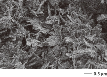
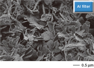
Specimen:Iron rust. Accelerating voltage:1kV, Detector:SED
NEW FUNCTION 2
Automatic large area acquisition
Montage
Montage images, which cover large areas with automatic acquisition of each area and correction of positional area shift, can be acquired simply by specifying the areas and acquisition conditions. Applying digital zoom to the acquired montage images displays detailed information about the specimen. In addition, montage elemental maps using EDS can simultaneously be obtained, enabling automatic acquisition of a great deal of information without the help of an operator.

NEW FUNCTION 3
Specimen Exchange System
Applicable to specimens of all sizes, as well as optical imaging
Draw-out system suitable for large-specimen exchange is employed as standard. It can also acquire optical image, searching for observation area and managing data can be faster and easier. Use of optional Specimen Exchange System enables fast and clean specimen loading/unloading.
