FEATURE
Enabling seamless observation and analysis
Further strengthened FIB processing capabilities. Greatly improved SEM imaging by a new optical column. Improved operability with linkage capabilities.
FIB : Further strengthened processing capabilities
- ・Enhanced control system
The vector scan system allows for smooth processing of an arbitrary shape.
Simple 3D observation & analysis is enabled. - ・Large ion beam current up to 90 nA
High-speed processing of the specimen is enabled.
SEM : Improved imaging performance
- ・High resolution at low accelerating voltage
Combination of a hybrid conical objective lens and GENTLEBEAM™ achieves high resolution at low accelerating voltage (1.6 nm at 1 kV). - ・Acquisition of a variety of images
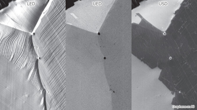
Newly added UED & USD detectors enable acquisition of a variet y of SEM images that contain information on properties, chemical compositions and crystal structures. - ・High resolution at large probe current
Combination of an “in-lens Schottky electron gun” and an aperture angle control lens(ACL) maintains high resolution at large probe current, allowin g for fast analysis.
3D observation and analysis
- ・Stable automatic measurement
The Slice and View system (standard component of the JIB-4700F) enables automatic repetition of processing, observation and analysis. - ・Easy reconstruction
After the repeated steps, a 3D reconstructed image is acquired by the 3D-reconstruction software (IB-67020STKV)* from the serially-acquired data.
APPLICATION
Versatile solutions offered by FIB-SEM
Cross section specimen preparation, TEM specimen preparation, 3D observation, 3D EDS analysis, 3D EDSD analysis, Processing of fine shapes
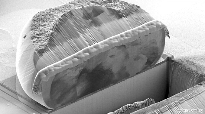
Cross Section
The JIB-4700F Multi Beam System allows for seamless operations from protective-film preparation, specimen milling to cross section observation and analysis. The FIB column enables processing with a largecurrent Ga ion beam (up to 90 nA). This largecurrent processing is particularly effective for a large-area specimen preparation.

TEM Sample
The combination of the JIB-4700F and the manipulator system allows for smooth TEM specimen preparation. The use of the backscattered electron detector enables you to observe the progress of FIB milling with a high-resolution SEM image. The JIB-4700F enhances operation efficiency, such as detection of final milling end-point for preparation of TEM specimens.

-
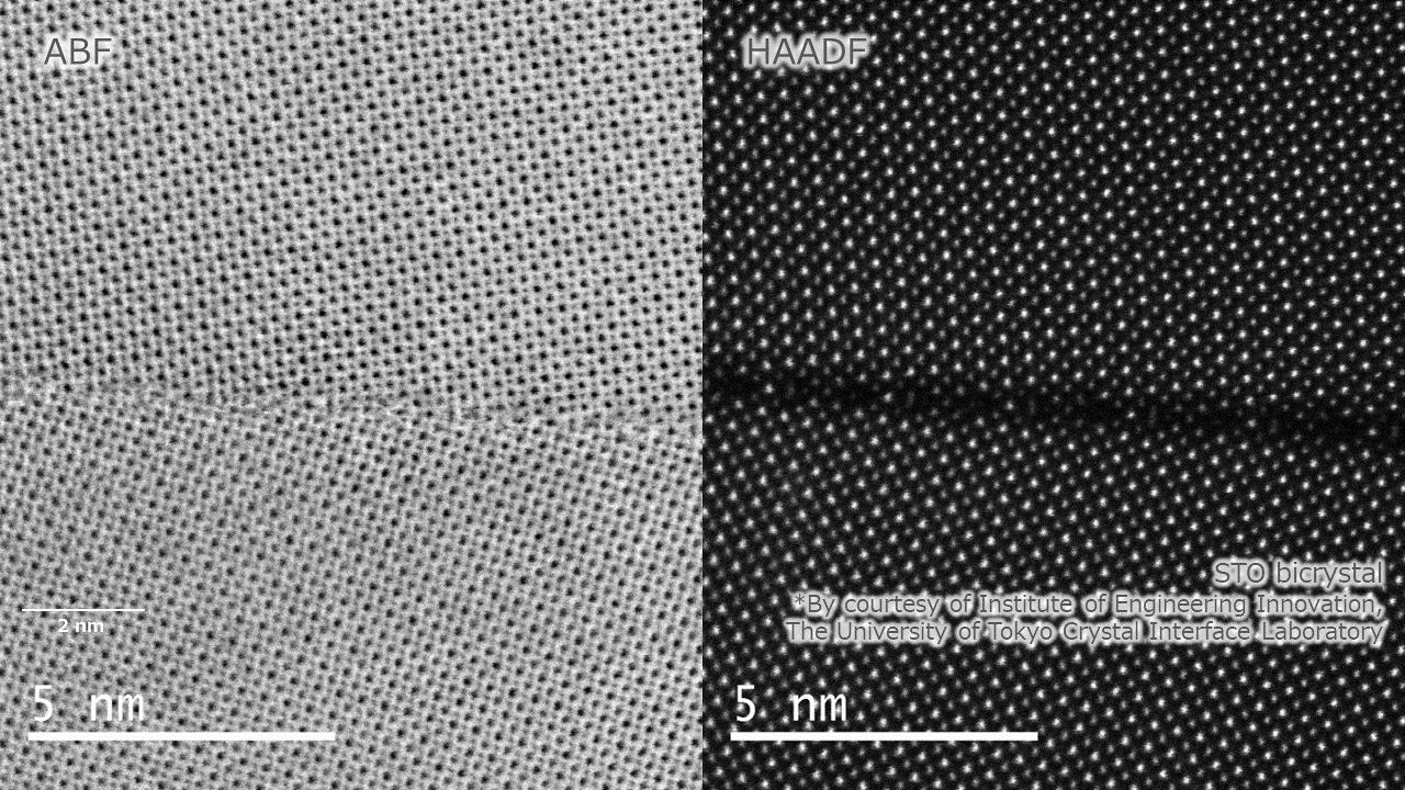
Atomic resolution STEM images acquired by JEM-ARM200F NEOARM
High quality TEM specimen prepared by low energy Ar ion beam polishing with IonSlicer™
-
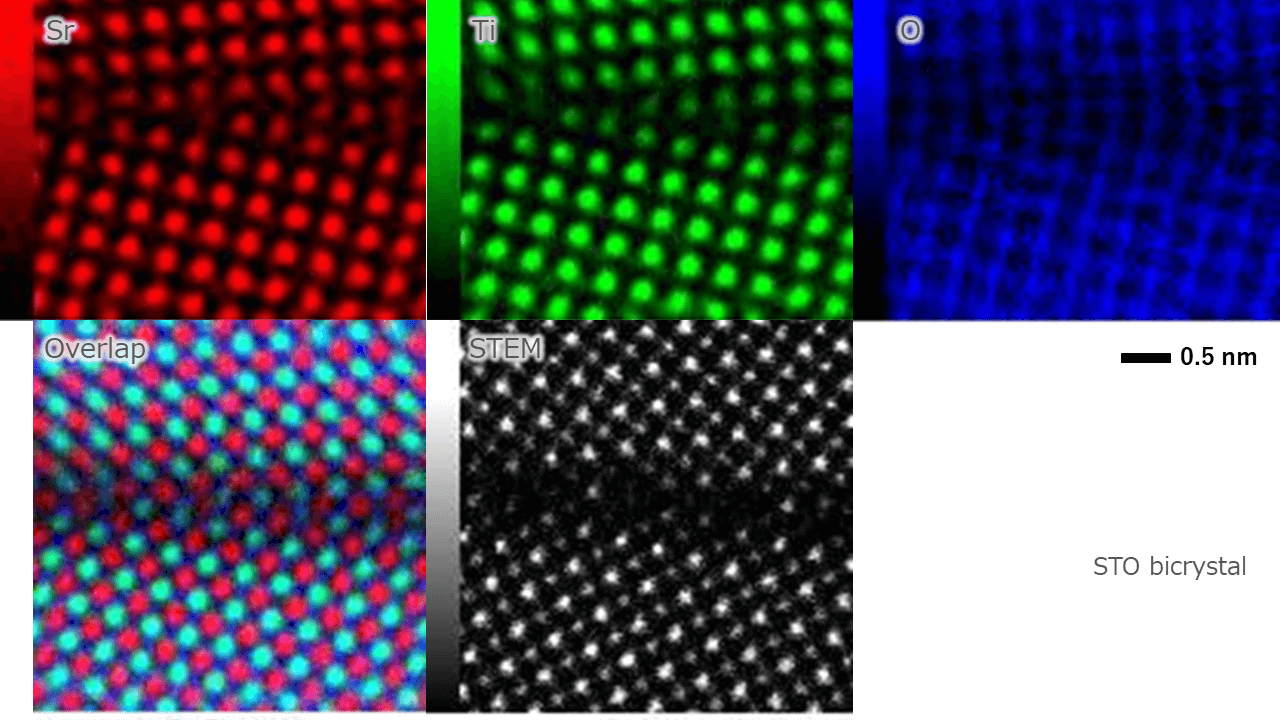
Atomic resolution EDS maps acquired by JEM-ARM200F NEOARM
High quality TEM specimen prepared by low energy Ar ion beam polishing with IonSlicer™
3D-EDS Analysis
3D-EDS* enables automatic serial steps of both FIB milling with an incident ion beam normal to the specimen surface and EDS analysis using a SEM probe.High resolution at large probe current allowin for fast EDS analysis.

3D-EBSD Analysis
An EBSD detector*, which is optimally placed,enables processing and analysis with no stage movement. This feature provides high positional accuracy of data acquisition with a shortened time.