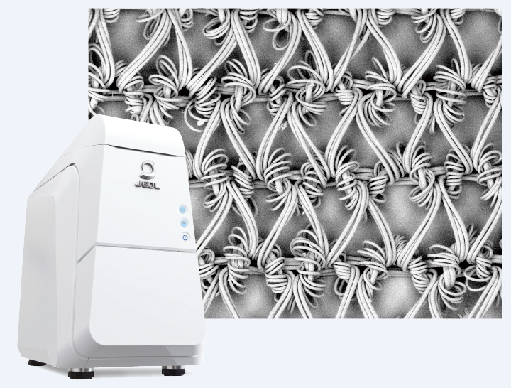
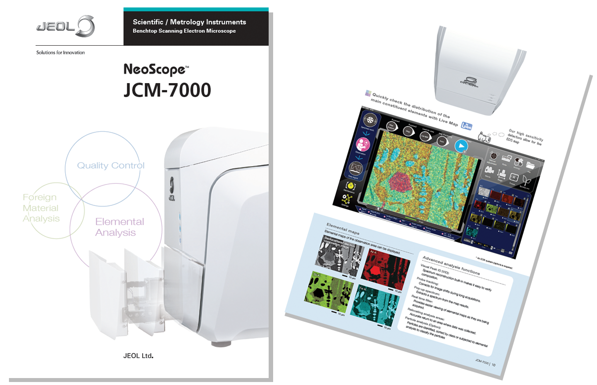
Easy-to-use SEM/EDS just for anyone!
JCM-7000 NeoScope™ Benchtop SEM
Brochure Download
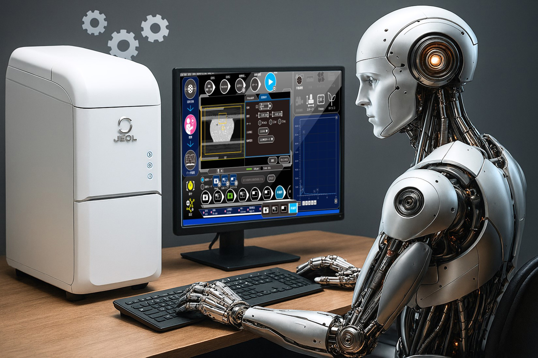
Solving the Shortage of SEM Operators!
- Neo Action
"Neo Action" enables the SEM to automatically queue up multiple measurement areas and define acquisition parameters such as: incident voltage, signal and adjusting the image. It automates routine tasks from data acquisition to reporting. If you are struggling with a shortage of SEM operators or busy multitasking, this is an essential function!
Continuous acquisition of structures of the same size
Shooting and analyzing at different magnifications within the same field of view
Comparing images and analysis with different incident voltages
Seamless transition from Optical to SEM imaging!
- Zeromag & Low-Vacuum Mode
An optical image is automatically acquired when the sample is inserted. Search for the field of view on the optical image, then zoom in on the target to automatically switch to an SEM image. Moving to the observation position is easy for quick SEM image acquisition with a minimal number of steps.
Even after switching to the SEM image, samples can be observed without pretreatment thanks to the standard low-vacuum mode, which also supports non-conductive and moisture-containing samples.
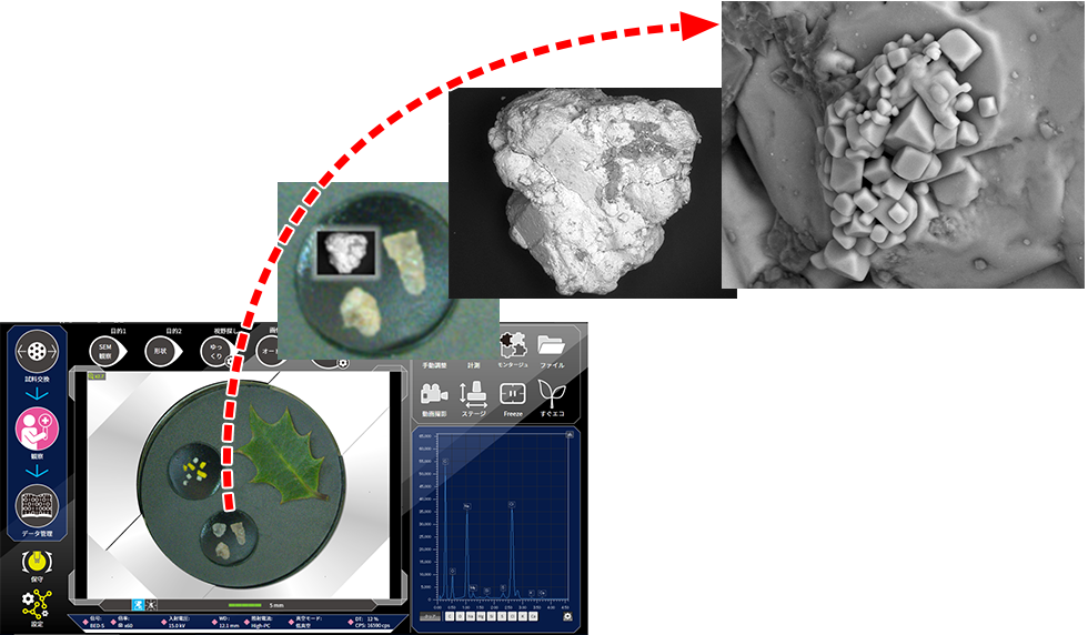
Specimen: Salt
Seamless transition from SEM imaging to EDS Analysis
- Live Analysis & Live Map
With Live Analysis, SEM observation and EDS analysis are no longer separate steps. The X-ray spectrum with the main constituent elements are displayed in Real Time on the observation screen. The JCM-7000 also includes Live Map to view the spatial distribution of the elements in Real Time. Live Map increases the probability of finding the elements of interest as well as detecting unexpected elements.
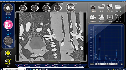
Screening while performing observation with Live Analysis
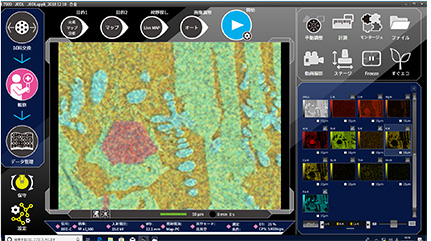
Quickly check the element distribution with Live Map
When 2D images are not enough
- Live 3D
The high-sensitivity 4-segmented backscattered electron detector enables 2-pane viewing of a SEM image and a 3D image using Live 3D function. In addition to instantaneous shape determination for samples with complex topographies, depth information can also be acquired.
Specimen: Coin
Images obtained with a 4-segmented backscattered electron detector

Live 3D image
Applications
Metal
For conductive metal specimens, observation of surface details using the secondary
electron image can be performed without coating.
With the JCM-7000, details of ductile or brittle fracture can be analyzed, including surface morphology of
the fracture, elemental analysis of materials present at the starting point of a fracture, and
identification of inclusions in metal.
Ductile or brittle fracture on glass
For the ductile or brittle fracture on transparent glass or plastics, it is
difficult to confirm its top-surface state with an optical microscope.
Observation with the SEM makes it easy to find the starting point of a fracture and observe the detailed
surface morphology.
Printed circuit board
Low-vacuum mode is suitable for a printed circuit board (composite material). Owing
to this mode, SEM observation and analysis can be performed without adding a conductive coating.
The Live 3D function enables an SEM image (BEI, shadow) and a live 3D surface reconstructed image to be
displayed simultaneously.
Fibers
For fibers with complex structure, adding a conducting coating is difficult. Low-vacuum mode makes it easy to perform morphological observation as well as analysis of foreign materials.
Food
Low-vacuum mode is effective for observation and analysis of food, which contains a
lot of water or fats.
In particular for specimens that are susceptible to heat, the use of an LV cooling holder (option) allows
for observation and analysis of the food specimen uncoated while preserving its structure.
Asbestos
SEM/EDS enables determination of the presence or absence of asbestos in building
materials by combining the results of morphological observation and compositional (elemental) analysis.
The Live Analysis function makes it possible to check the spectrum while observing the SEM images. This
allows accurate, efficient judgment about the presence of asbestos when fibers are discovered.
Powder
It can be difficult to identify the type of powder adhered to a component simply by
the color.
With SEM, it is possible to identify the elements* as well as confirm details about the powder's morphology,
particle diameter, and adhesion.
Brochure Download
JCM-7000 NeoScope™
- Improved work efficiency with JCM-7000
- Easy Operation with JCM-7000
- Seamless transition from Optical to SEM imaging!
- Seamless transition from SEM imaging to EDS Analysis
- Simple report creation and data management
- Options to extend SEM capabilities
- Discover a New World with JCM-7000
- Easy maintenance
- Peripherals
About Us
JEOL Ltd.
Since its foundation in 1949, JEOL has been committed to the development of
cutting-edge scientific and metrology instruments, industrial and medical equipment.
Today, many of our
products are used throughout the world and we are highly regarded as a truly global company.
Aiming to
be
a 'top niche company that supports science and technology around the world', we will continue to respond
precisely to the increasingly sophisticated and diverse needs of our customers.