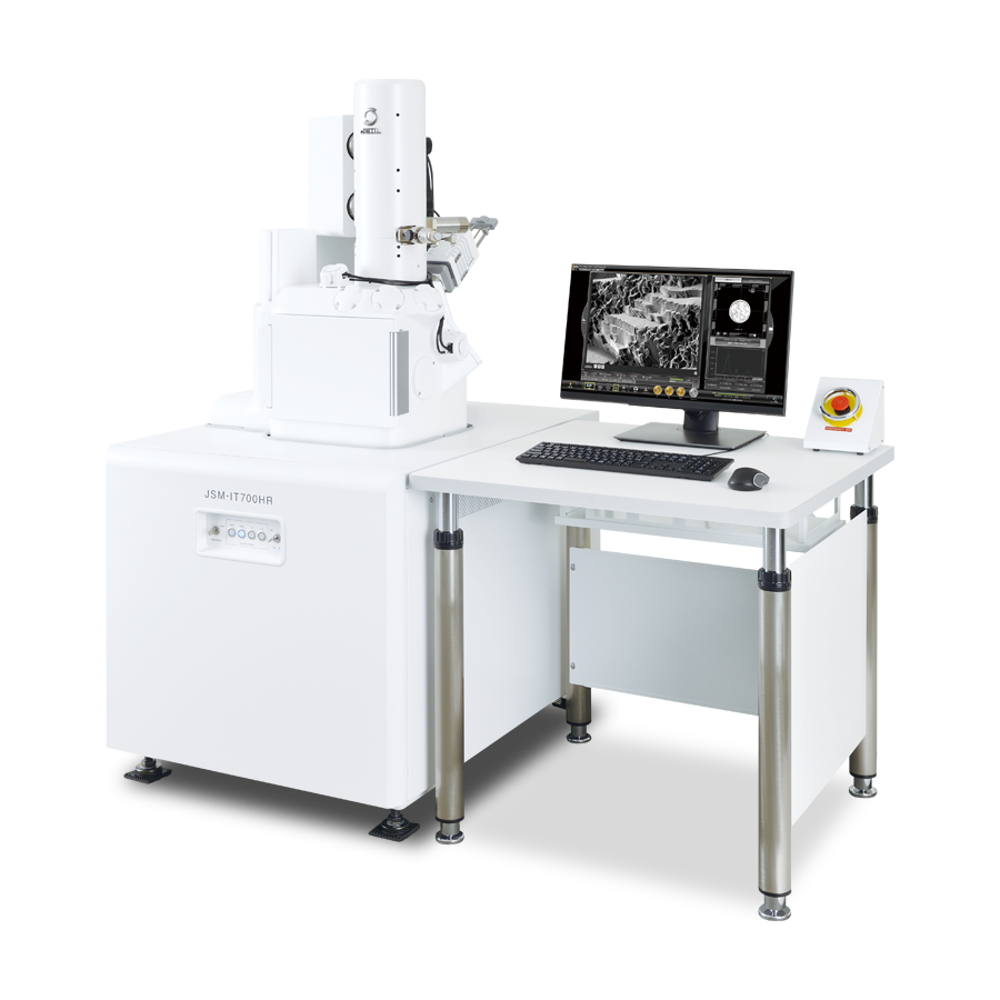
JSM-IT700HR
InTouchScope™ Scanning
Electron Microscope
SEM- Essential in Daily Lab Operation JSM-IT700HR Makes it Easy.
Features
Nano-scaled materials are driving the current technological breakthroughs and their observation and analysis is facilitated by a new and innovative SEM, JSM-IT700HR.
Its new electron gun with spatial resolution of 1 nm and the largest probe current of 300 nA, combined with an exceptionally userfriendly software interface significantly simplifies observation and analysis in SEM.
The compact instrument design also features a large specimen chamber with multiple accessory ports as well as EDS integration.
JSM-IT700HR Advanced SEM, Powerful and Simple to Use.
Introduction
♦Click the "replay" button in the box above, and the movie will start (about 1.5 min.) ♦
Zeromag & Autofunctions
Magnify optical image, seamleass transition to the SEM image
♦Click the "replay" button in the box above, and the movie will start (about 1.5 min.) ♦

Zeromag is designed to link the holder graphic or optical image※ with the SEM image.
Using Zeromag, field searching is easy.
Stage Navigation System (SNS) is necessary to display an optical image.
Integrated EDS & Live Analysis
Integration of observation and analysis
♦Click the "replay" button in the box above, and the movie will start (about 2 min.) ♦

EDS analysis directly on the SEM observation screen for seamless transition from observation to analysis. Moreover, Live Analysis provides real-time monitoring of the spectra for characteristic X-rays.
SMILE VIEW™ Lab
Fast and flexible report generation
♦Click the "replay" button in the box above, and the movie will start (about 1 min.) ♦

SMILE VIEW™ Lab is a JEOL original data management tool, which links the optical image, SEM image and EDS analysis results.
With one click, a report can be generated easily after the measurement.
An off-line version of the software*1 is available to free up the SEM and enhance productivity.
Off-line data analysis software (option) is required.
Need to install Microsoft®Office.
Display the depth of signal
♦Click the "replay" button in the box above, and the movie will start (about 1 min.)♦

A new function for displaying the generation depth of signal is built-in. Observing the analysis depth on the specimen is very effective for understanding the elemental results generated.
Auto Beam Alignment (ABA)
♦Click the "replay" button in the box above, and the movie will start (about 1.5 min.) ♦

This function automatically adjusts the optical axis of the electron beam.
Large area observation and analysis with montage function


Montage result: 6×6 (Left: Backscattered electron image Right: Cu element map)
Specimen: Flat milled section of brass screw※, Accelerating voltage: 20 kV, Low vaccum mode (20 Pa), Imaging area: 6.4 mm × 4.8 mm
Flat milling fabriation was performed by IB-19530CP after mechanical polishing.
Montage is a function to connect all images in a large area as one high-definition image.
This function is very useful for acquiring detailed information over a large area.
Catalogue Download
JSM-IT700HR InTouchScope(TM) Scanning Electron Microscope
Application
Application JSM-IT700HR
Introducing Cryo Scanning Electron Microscopy
Expanding the microscopic world through JSM-IT700HR
Nanomaterials

Carbon nanotube
Accelerating voltage: 2 kV, Signal: Secondary electrons,
Magnification: ×100,000
Observation at low accelerating voltage clearly reveals the surface structure.
Catalyst Pt on carbon

Electronic products

Fractured surface of ceramic capacitor
Accelerating voltage: 5 kV, Signal: Backscattered electrons,
Magnification: ×1,000 (left) ×10,000 (right)

CP-milled section of semiconductor SRAM
Accelerating voltage: 5 kV, Signal: Backscattered electrons, Magnification: ×60,000 (left, right)

CP is an instrument for preparing a cross section of a specimen using a broad Ar ionbeam and shield plate. In recent years, CP has been widely used to prepare cross sections of metal, ceramics, plastic,and other materials.
Metals
Large area montage analysis

Fracture surface of stainless
Accelerating voltage: 15 kV, Signal: Secondary electrons,
Magnification: ×500, Montage result: 13×6
By observing the entire area of a fracture surface, a detailed analysis of the fracture mechanism can be made. In this specimen, typical fatigue failure, such as the striation pattern and dimple microvoids, are observed.
Elemental analysis: EDS map

CP-milled section of precision cutting blade
Accelerating voltage: 15 kV, Signal: Backscattered electrons (left) EDS map (right), Magnification: ×3,000
Using overlay map, the distribution of heavy metal elements in the precision cutting blade is made clear.
High magnification EBSD analysis

CP-milled section of stainless wire along the longitudinal direction
Image Quality Map(left), Phase map image(right)

EBSD map image
(direction: Direction 3)
Accelerating voltage: 10 kV, Probe current: 5 nA, Magnification: ×10,000
Soft materials

Carbon black in the rubber
Accelerating voltage: 15 kV
Signal: Secondary electrons
Magnification: ×20,000

Plastic glove
Accelerating voltage: 5 kV,
Signal: Low vacuum backscattered electrons
Magnification: ×30,000

Membrane on a chicken eggshell
Accelerating voltage: 5 kV,
Signal: Low-vacuum secondary electrons
Magnification: ×500

Low-vacuum mode
Low vacuum mode allows for observation of non-conductive materials without treatment. Evacuation at the objective lens improves image quality in low vacuum mode.
Food

Ice cream
Accelerating voltage: 7 kV, Signal: Low vacuum backscattered electrons, Magnification: ×300 (left) ×30,000 (right)

Fat globules and muscle fiber of chicken
Accelerating voltage: 10 kV
Signal: Low-vacuum backscattered electrons
Magnification: ×300

LV cryo-holder*1
LV cryo-holder keeps a specimen frozen without water loss.
A hydrous specimen like food can be observed. It is possible to visualize the texture by understanding the size of ice and the diameter of muscle fibers.
*1 Optional
Biology

E. coli and T4 phage
Accelerating volage: 2.5 kV
Signal: Secondary electrons
Magnification: ×25,000

Accelerating volage: 2.5 kV
Signal: Secondary electrons
Magnification: ×80,000

Mitochondria of mouse kidney
Accelerating voltage: 2.5 kV
Signal: Secondary electrons.
Magnification: ×50,000

JFD-320 Freeze Drying Device*2
This freeze drying device minimizes the effect of surface tension, suitable for drying hydrous specimens.
Specimen preparation of E. coli and T4 phage: Critical point drying after Glutaraldehyde and OsO4 treatment.
Specimen preparation of mouse mitochondria: Freeze drying after OsO4 maceration treatment.
* 2 Optional
Related Products
Related Products
IB-19520CCP CROSS SECTION POLISHER™
Thermal damage can be reduced by cooling the specimen with liquid nitrogen during processing.
Designed to suppress the consumption of liquid nitrogen, allowing long cooling periods.
Rapid cooling of the specimen while immersed in liquid nitrogen. Return to room temperature. Designed to allow parts to be detached.
Incorporates a mechanism to allow the process from polishing to observation to be performed without exposing the sample to the air.

IB-19530CP CROSS SECTION POLISHER™
The IB-19530CP features an innovatively designed, multi-purpose stage to fulfill increasingly diversified market needs and realize multi-functionality by different kinds of functional holders. The multi-purpose stage combined with specialized functional holders allows the user to perform various functions such as planar surface milling and polishing, sputter coating as well as more traditional cross-section ion milling.
More Info
Are you a medical professional or personnel engaged in medical care?
No
Please be reminded that these pages are not intended to provide the general public with information about the products.